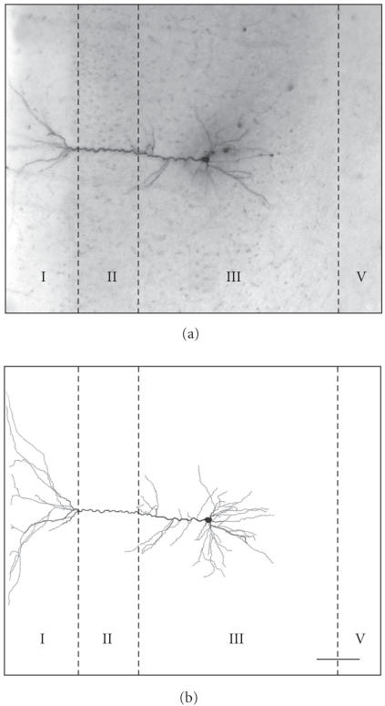
Figure 2.
Example of an intracellularly labeled and reconstructed pyramidal neuron in the PFC of a control rat. (a) Photomicrograph of an intracellularly labeled pyramidal neuron in layer III of the prelimbic subarea (left hemisphere). (b) Line drawing of the neuron shown in (a) (reconstruction with NeuroLucida). The relative position of the pyramidal cell is shown by lines indicating the cortical layers (I–V). Scale bar: 100 μm.

