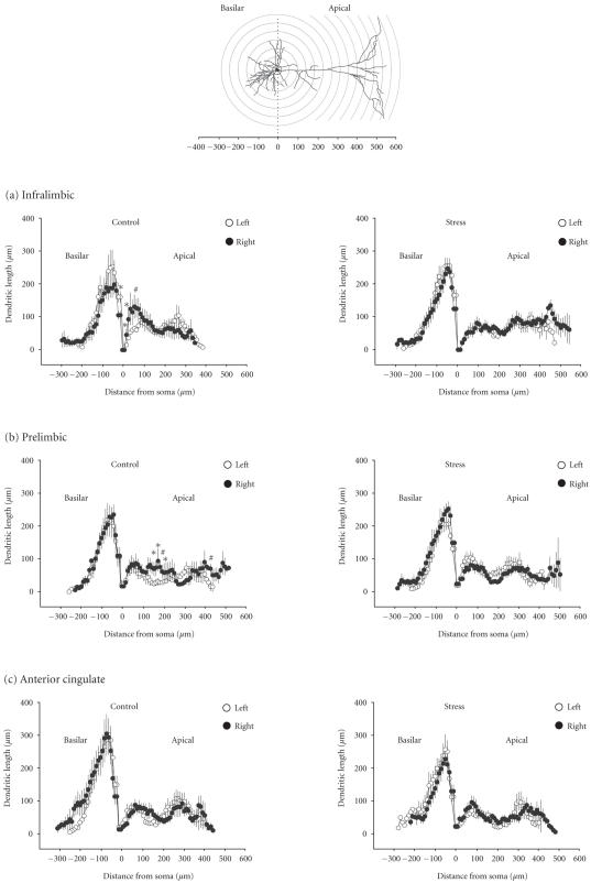
Figure 3.
Sholl analysis of dendrites on pyramidal cells in the left (open circles) and the right hemisphere (closed circles) of controls (left panel) and stressed rats (right panel). Basilar dendrites are plotted to the left and apical dendrites to the right as a function of the distance from the soma center (0). The schematic drawing in the top panel illustrates the Sholl circles that correspond to the distances from soma depicted in (a) infralimbic, (b) prelimbic, and (c) anterior cingulate. Data points represent the sum of all dendrites detected at the respective distance (radius) from the soma center set as zero (mean ± SEM). Symbols indicate significant differences within each 10 μm ring determined by ANOVA with Bonferroni's post hoc test (∗P < .05, #P < .01).

