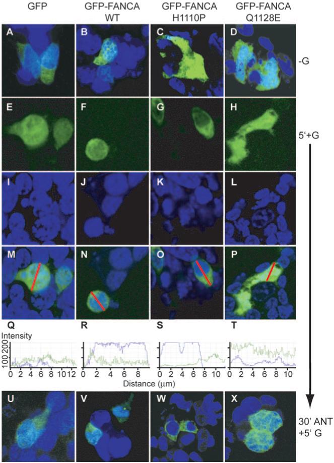
FIG. 6.

Point mutations block GnRH-induced nucleocytoplasmic shuttling of FANCA. Cells were transfected with either GFP-empty vector (GFP) or GFP-FANCA (WT) or the point-mutated GFP-FANCA mutants (H1110P or Q1128E) and then left untreated or treated with 100 nm GnRH for 5 min. Cells were fixed before staining nuclei with Torpro-3. A-D, Merged untreated (−G). GFP-(E-H) and Torpro-3-stained nuclei (I-L) are merged (M-P) and demonstrate the differential locations of untagged and tagged GFP after addition of GnRH for 5 min (5′+G). A line was drawn through the merged cell (M-P), and the intensity of the fluorescence, ranging from zero to a maximum of 256, was measured by calculating how many pixels hit the line for each individual channel, GFP in green, Topro-3 in blue, and are shown graphically in Q-T. U-W, Localization of untagged and GFP-tagged proteins after 30 min pretreatment with 1 μm of GnRHR antagonist Cetrorelix (30′ANT), then 5′+G. These are representative examples of images taken from at least three separate experiments.
