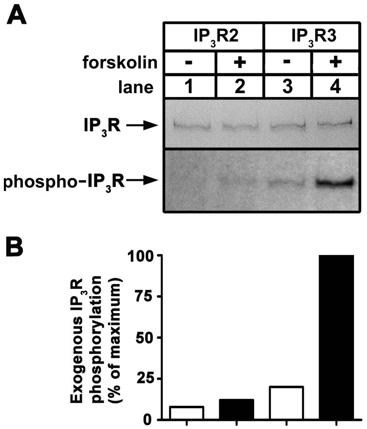
Figure 1. Phosphorylation of IP3Rs in intact cells.
(A) [32P]Pi-labeled HEK cells transfected with cDNAs encoding wild-type IP3R2 (lanes 1-2), or IP3R3 (lanes 3-4) were exposed to 10 μM forskolin for 5 min as indicated, and IP3Rs were immunoprecipitated with CT2 or CT3, electrophoresed and assessed for Coomassie blue staining capacity (upper panel) and associated radioactivity (lower panel). Negligible staining and radioactivity was obtained from control (vector-transfected) cells (not shown) [14, 18]. (B) Histogram of the data presented in A. Exogenous IP3R phosphorylation was calculated by subtracting the negligible radioactivity obtained from control cells from that in lanes 1-4 and is expressed as a percentage of the radioactivity associated with IP3R3 in the presence of forskolin (lane 4). Data shown are representative of three independent experiments.

