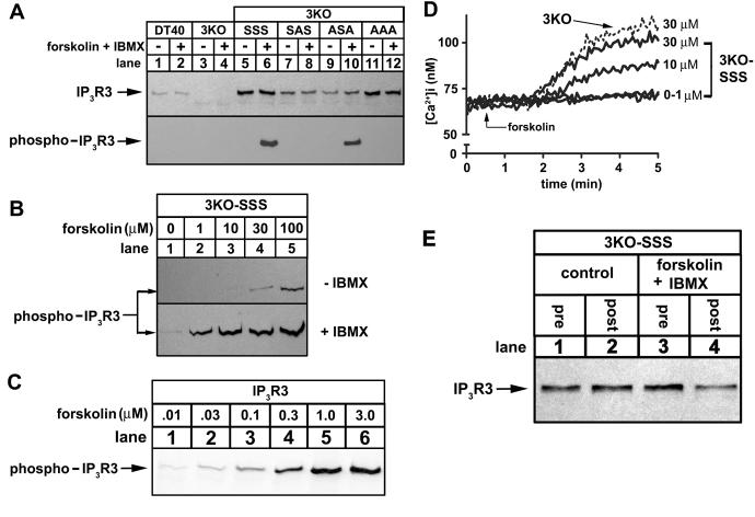
Figure 3. Phosphorylation of IP3R3s expressed in 3KO cells.
(A) DT40 cells (lanes 1-2), 3KO cells (lanes 3-4), or IP3R3-expressing 3KO cells (lanes 5-12) were exposed to 1 μM forskolin plus 200 μM IBMX for 10 min as indicated, lysates were prepared, and IP3R3s phosphorylated at S934 were immunoprecipitated with anti-pS934. Total IP3R3 in lysates prior to immunoprecipitation (upper panel) or phosphorylated IP3R3 in immunoprecipitates (lower panel) were assessed in immunoblots with TL3. (B) 3KO-SSS cells were exposed to 0-100 μM forskolin in the absence (upper panel) or presence (lower panel) of 200 μM IBMX for 10 min, and IP3R3 phosphorylation was assessed as in A. (C) IP3R3SSS-expressing HEK cells were exposed to 0.01-3.0 μM forskolin for 10 min and IP3R3 phosphorylation was assessed using anti-pS934 in immunoblots of cell lysates. (D) Traces of typical Ca2+ responses to forskolin (added at t = 0.5 min) in 3KO cells (dashed line) and 3KO-SSS cells (solid lines). (E) 3KO-SSS cells were incubated with or without 1μM forskolin plus 200 μM IBMX for 10 min as indicated and levels of IP3R3 in cell lysates prior to (pre) and following (post) immunoprecipitation of phosphorylated IP3R3 with anti-pS934 were assessed in immunoblots with TL3. The lack of difference in signal intensity in control lysates pre and post immunoprecipitation (lanes 1 and 2) shows that IP3R3SSS is not significantly phosphorylated, whereas the difference in signal intensity in stimulated cells (lanes 3 and 4; a reduction of ∼50%) shows that at least 50% of IP3R3SSS is phosphorylated.

