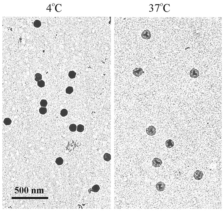
Figure 4.

Electron micrographs of HSV-1 C capsids bound to Formvar/carbon-coated electron microscope grids. Capsids were maintained at 4°C (left) or warmed to 37°C (right) for 2 hrs and stained with uranyl acetate. Note that DNA loss, as evidenced by a lighter image, is observed only in capsids heated at 37°C.
