
Fig. 5.
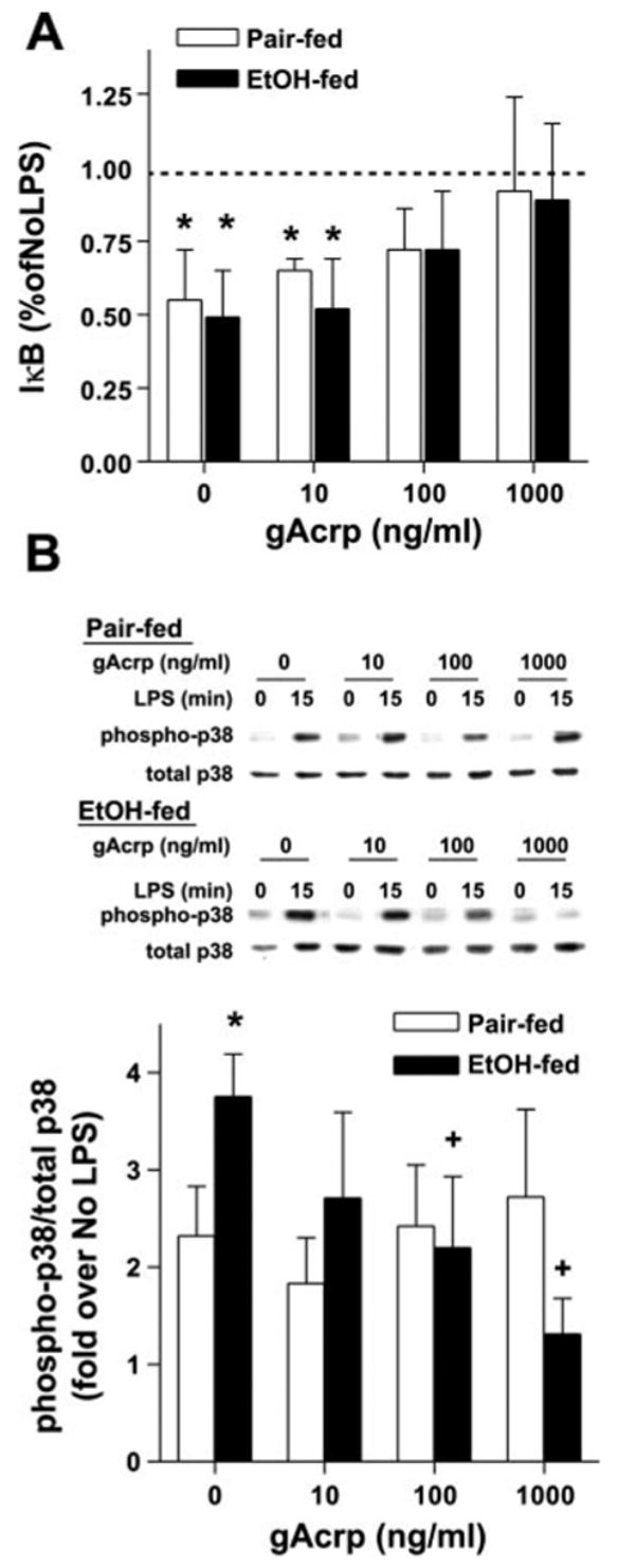
Effect of gAcrp on LPS-stimulated IκB degradation (A) and p38 MAPK phosphorylation (B) in Kupffer cells from EtOH-fed rats. Kupffer cells from pair- and EtOH-fed rats were cultured for 16 h with or without 10–1,000 ng/ml gAcrp. A: cells were then stimulated or not with 100 ng/ml LPS for 30 min to measure IκB degradation. Values represent means ± SE; n = 5. *P < 0.05 compared with cells not treated with LPS in the same diet group. B: cells were then stimulated or not with 100 ng/ml LPS for 15 min to measure phosphorylation of p38. Values represent means ± SE; n = 4. *P < 0.05 compared with pair-fed rats at each concentration of gAcrp; +P < 0.05 compared with cells not treated with gAcrp in the same diet group.