
Figure 5. Cell Intrinsic Block to B Cell Development in Sfpi1BN/BN mice.
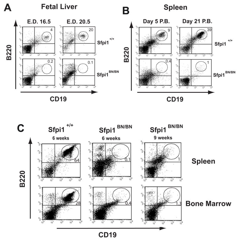
(A, B) Flow cytometric analysis of single-cell suspensions from either fetal livers or spleens of wild-type and Sfpi1BN/BN mice at the indicated age. Isolated spleen cells were analyzed by flow cytometry using gating for size and granularity and antibodies to the indicated cell surface markers. Numbers indicate percentage of gated cells in the indicated region. E.D. = emybryonic day, P.B. = post birth. (C) Flow cytometric analysis of single-cell suspensions from spleens or bone marrow of recipient mice transplanted with wild-type or Sfpi1BN/BN fetal liver cells. Fetal liver cells were isolated from wild-type or Sfpi1BN/BN mice and transplanted by tail vein injection into sublethally irradiated Rag2−/− Il2rg−/−recipient mice. Analysis was performed after the indicated time following transplantation.