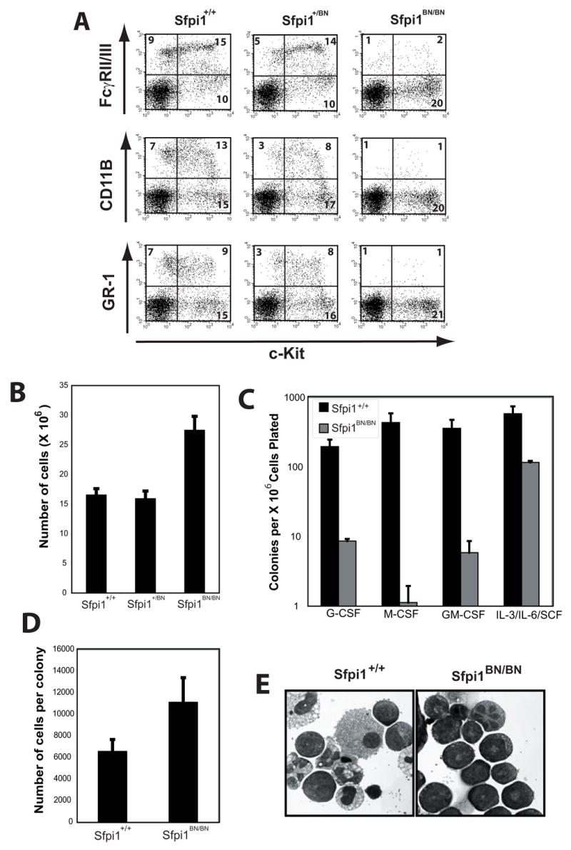
Figure 6. – Fetal Liver Myelopoiesis is Severely Reduced in Sfpi1BN/BN Mice.

(A) Flow cytometric analysis of single-cell suspensions from day 14.5 fetal liver cells. The cell surface markers analyzed are shown. Numbers indicate the percentage of total cells. (B) Total number of cells per fetal liver of wild-type, heterozygous, and Sfpi1BN/BN mice. (C) Colony assays from fetal liver progenitors isolated from wild-type and Sfpi1BN/BN mice. Fetal liver cells were isolated from the indicated mouse and plated at various concentrations in methylcellulose with the indicated cytokines. Colonies were scored following 7 days of culture. Error bars represent the standard error of a minimum of three plates. (D) Calculated cells per colony of either wild-type or Sfpi1BN/BN fetal liver progenitors. Cells in methylcellulose cultures from (C) were washed and counted. (E) Cytospins of cells from methylcellulose plates described in (C and D).
