
Figure 3. aff-1 Is Required for AC Fusion prior to Cytoplasmic Mixing.
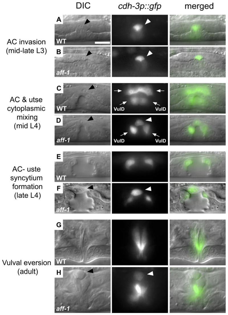
AC, arrowhead; utse syncytium, arrows.
Nomarski (left) fluorescence (center) and overlaid (right) images of vulval-uterine area in critical intermediates of AC development during L3 to adult.
(A) AC invasion in the L3 stage was detected by a cadherin promoter driving GFP expression (cdh-3p::GFP; [Sherwood and Sternberg, 2003]).
(B) In aff-1 mutant, AC invasion is normal.
(C) In wild-type, cytoplasmic mixing between AC and utse cells is detected by diffusion of the AC marker cdh-3p::GFP to the utse (arrows); VulD represents vulval ring “D.”
(D) In aff-1 mutant, cdh-3p::GFP retention in the AC demonstrates that cytoplasmic mixing does not occur (arrowhead).
(E) At the vulval “Christmas tree” stage, the AC and utse syncytium form a thin layer between vulva and uterus lumens in wild-type.
(F) In aff-1 mutant, this layer is not formed and the unfused AC lies at the vulva-uterus junction (arrowhead).
(G) Normal adult vulva after eversion.
(H) Unfused AC remains at the apex of the everted aff-1 vulva (arrowhead). All panels are at the same magnification; the scale bar corresponds to 5 μm.