
Figure 4. aff-1 Is Required for Fusion Events in Other Tissues.
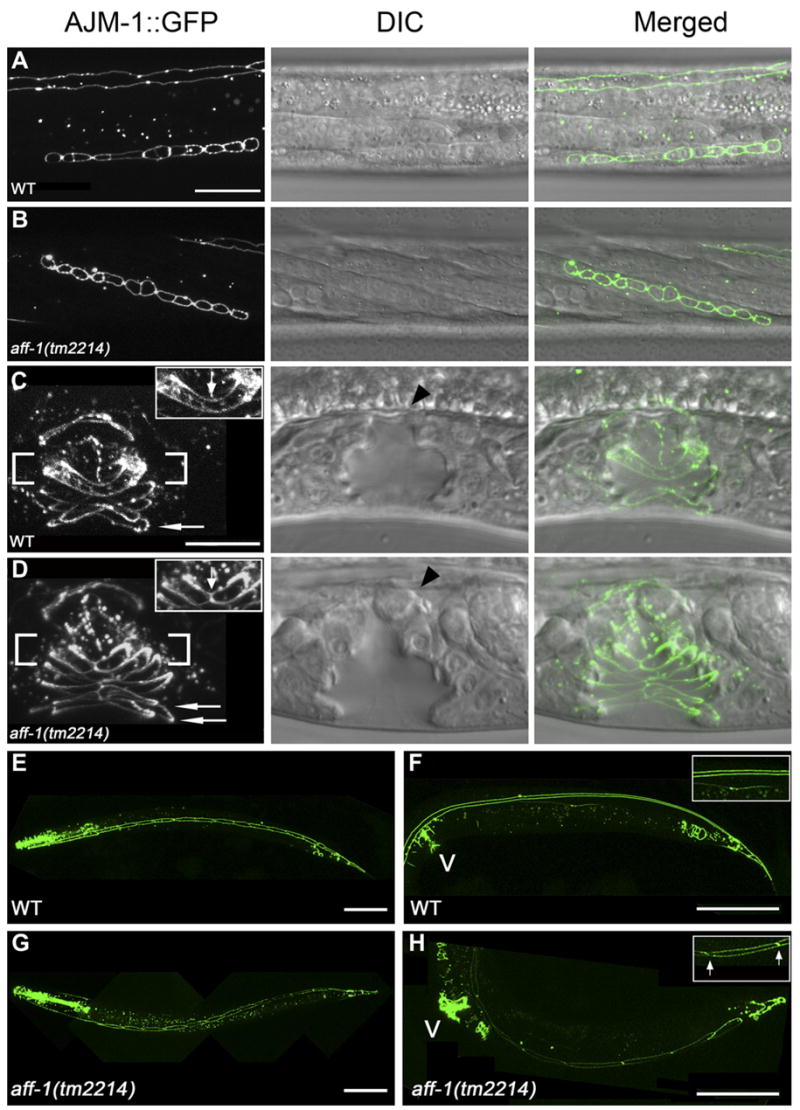
(A–D) Nomarski (center) and the corresponding fluorescence image in selected stages of vulval development of the apical junction marker AJM-1::GFP that marks epithelia cell borders.
(A) In wild-type worms, 12 primordial vulval cells are located at the ventral side at late L3 stage.
(B) A similar pattern in aff-1 mutants shows that aff-1 does not affect VPCs proliferation. In addition, the fusion of 3° fate VPCs to the epidermis is normal.
(C) Fusion of vulval cells results in the formation of vulval rings in wild-type (for example vulD ring, fusion marked with an arrow in inset). vulA represents a single ventral ring (arrow).
(D) In aff-1 deletion, the two D cells did not fuse; hence the D ring is unfused (arrow in insert). The A cells fail to fuse before ring formation and two vulA rings form instead of one (arrows).
(E–H) Selected stages of seam cell development in wild-type (E and F) and aff-1 mutant worms (G and H) examined by the AJM-1::GFP marker.
(E) In wild-type L3 stage, 16 seam cells are on each side of the body, separated by apical junctions (left view).
(F) During late L4/early adult, seams undergo cell fusion that results in a long syncytium marked by two parallel lines of AJM-1::GFP; see insert and top of (A).
(G) In aff-1 mutant, early seam development is similar to wild-type.
(H) During late L4, the seam syncytia did not form, so individual cells are detected and remained unfused in adults. Insert shows detail with unfused apical junctions, arrows.
(A, B) and (C, D) are panels with same magnification. The scale bars in (A) and (C) represent 10 μm. (E–H) Scale bar corresponds to 50 μm. V, vulva.