
Fig. 4.
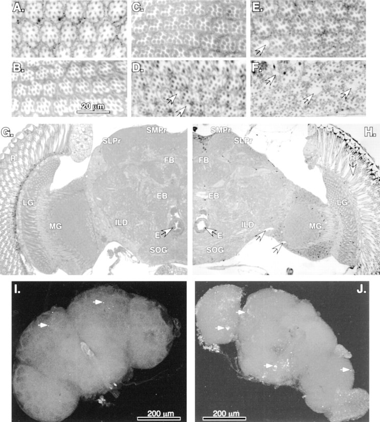
Neural degeneration and apoptosis inbchs mutants. A–F, Plastic-embedded tangential sections (1 μm) of adult retina stained with methylene blue. Minor differences in early ommatidial morphology are attributable to the depth of each individual section and position within a given eye. A, Retinal morphology of Canton Scontrols at 1 d of age is very similar to that ofbchs3/Df(2L)clot7 (B) individuals of the same age.C, At 10 d wild-type controls demonstrate the mature architecture and cellular morphology ofDrosophila retina, whereas age-matchedbchs3/Df(2L)clot7 flies (D) show the first signs of degeneration (vacuoles noted by arrows). E, At 14 d of age the wild-type retinas appear healthy and intact, with only the rare development of small vacuoles (arrow).F, At 14 d the degeneration detected earlier inbchs3/Df(2L)clot7 mutant flies has progressed further. There is a substantial loss of ommatidial architecture and an enlargement of vacuoles (arrows).G, H, Frontal sections (1 μm) from 10-d-old adults taken at the same depth within the head, near the central complex, as noted by the esophagus (E, arrows) and fan-shaped (FB) and ellipsoid bodies (EB). G, A section taken from 10-d-old controls demonstrates the characteristic organization and size of mature Drosophila neural structures. H, Age-matched section from abchs3/Df(2L)clot7 fly shows the presence of retinal degeneration (arrow) as well as atrophy of laminal (LG) and medullary (MG) ganglia, inferior lateral deutocerebrum (ILD), superior lateral (SLPr) and medial (SMPr) protocerebrum, and subesophageal ganglia (SOG) structures of the CNS. Nuclei and background appear darker in this image because of slight image enhancement.I, J, In situ TUNEL analysis of 2-week-old brains. In wild-type brains (I) the arrows indicate the limited number of cells undergoing apoptosis [402CyO/Df(2L)clot7; n = 10]. bchs null brains (J) have extensive TUNEL labeling, indicating significant numbers of cells undergoing apoptosis in most cortical regions of the CNS [Df(2L)dsf3/Df(2L)clot7; n = 7].