
Fig. 5.
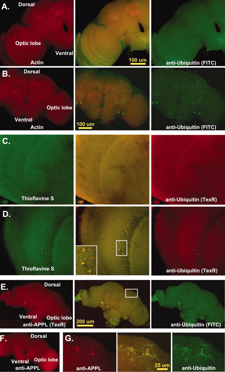
CNS protein aggregate formation.A–G, Single 0.2 μm transverse optical sections taken at least 5 μm into adult CNS. A, Optical section from an 11-d-old wild-type adult brain (n = 27) stained for actin (phalloidin-TRITC, Texas Red) and ubiquitin (FITC). Normal flies have a consistent pattern of actin and ubiquitin proteins within the CNS. Actin is enriched in neural projections and regions of synaptic innervation, whereas ubiquitin is stained uniformly throughout the entire CNS. B, Age-matched bchs null brains (n = 53) have deposits of ubiquitin in the CNS, mainly in regions of neuropil. C, Confocal images of 10-d-old wild-type controls (n = 20) stained for thioflavine S (green) and anti-ubiquitin (TexR).D, Identical images from age-matchedbchs3/Df(2L)clot7 flies (n = 15) stained with thioflavine S and for ubiquitin aggregates. E, Confocal sections from 11-d-old Df(2L)dsf3/Df(2L)clot7 adult brain (n = 22) stained for APPL (TexR) and ubiquitin (FITC).Drosophila APPL colocalizes with ubiquitin deposits in the CNS. F, APPL aggregates do not form in 2-week-old wild-type controls (n = 12). G,Inset enlargement (through portions of the mushroom body) shows the close association of ubiquitin and APPL in protein aggregates.