
Figure 1.
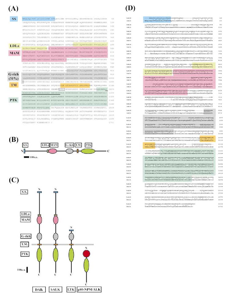
Molecular characterization of DAlk. (A) Protein sequence of DAlk. The protein sequence for DAlk is shown. Though not shown here, an in-frame upstream STOP codon precedes the initial methionine residue. The termination STOP codon is indicated by an asterisk (*). The signal sequence, LDLa, MAM, G-rich, transmembrane and PTK domains are shaded, and the consensus GxGxxG and AxK in the ATP binding site is underlined. The originally identified 64 amino acid PCR clone is double underlined. The putative IRS/SHC SH2 binding site (NPNY1170) is boxed. (B) DAlk schematic outline. (C) Schematic comparison of DAlk with the mammalian LTK/ALK family RTKs, ALK. Also shown is the p80-NPM/ALK protein product of the t(2;5) chromosomal translocation found in non-Hodgkins lymphoma. Structural conservation between DAlk, hALK and LTK include the presence of an intracellular PTK domain, an amino-terminal signal sequence, and an extracellular glycine-rich domain. Only DAlk and hALK share extracellular LDLa and MAM domains. (D) Alignment of the deduced amino acid sequences of Drosophila and human ALK. The signal sequence, LDLa, MAM, G-rich, transmembrane and PTK domains are shaded. The putative IRS/SHC SH2 binding site (NPNY1170) is boxed. The amino acid residues which are identical between the two species are marked by asterisks.