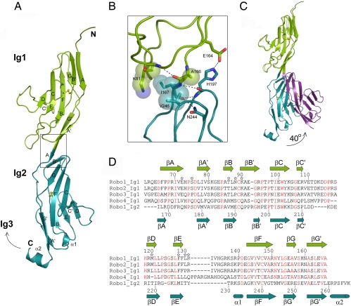
Fig. 1.
Structure of human Robo1 Ig1–2. (A) Ribbon diagram. The disulfide bridges are in yellow, and the box indicates the region highlighted in B. (B) Residues involved in interdomain contacts of the Ig1-Ig2 interface. (C) Ribbon diagram of the two Ig1–2 crystal forms showing the hinge movement of Ig2. (D) Sequence alignment of Ig1 domains of human Robo1, -2, -3, and -4 and of the Ig2 domain of human Robo1. Residue numbering is for Robo1 Ig1 (above) and Robo1 Ig2 (below). Slit2 D2-binding residues selected for mutagenesis are marked with a star, and residues strictly conserved between the Ig1 domain of human Robo1, -2, -3, and -4 are shown in red.

