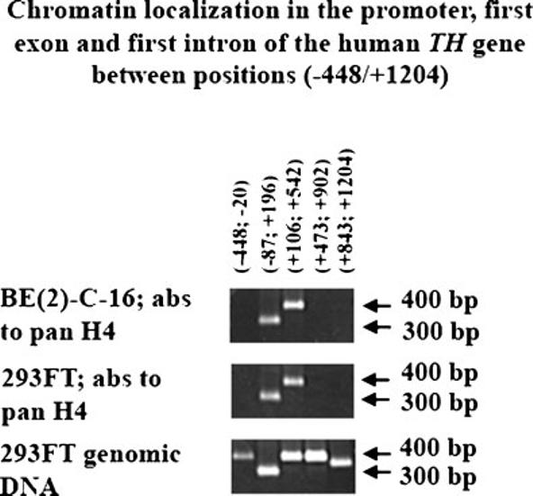
Fig. 4.

Chip analysis of chromatin localization in the promoter, first exon and first intron of the human TH gene between position (−448/+1,204). Antibodies (abs). Pan antibodies to H4 were used for Chip analysis of genomic DNA and chromatin complexes extracted from human neuroblastoma BE(2)-C-16 and human renal carcinoma 293FT cell lines (Materials and Methods). Five sets of sense and antisense primers were used for PCR analysis of Chip products (Table 1). The coordinates of the genomic DNA regions analyzed are reported on top of the first part. Two arrows depict 300 and 400 bp PCR products per each of the three parts (right hand-side). Chip analysis of genomic DNA and chromatin complexes extracted from human neuroblastoma BE(2)-C-16 cell line is shown in the top part, whereas the middle part shows the Chip analysis products of genomic DNA and chromatin complexes extracted from human renal carcinoma 293FT cell line. The lower part shows PCR products obtained for the five sets of sense and antisense primers using as template genomic DNA extracted from 293FT cell line.
