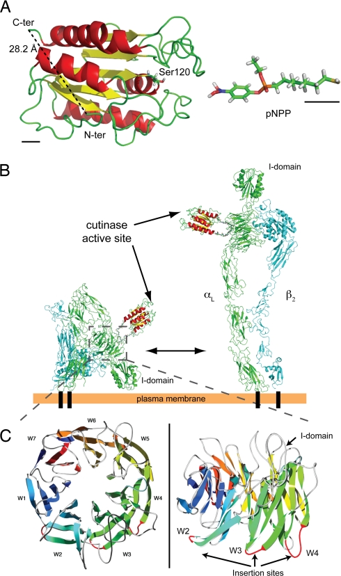
Fig. 1.
Insertion of cutinase into integrin αL. (A) Tertiary structure of cutinase (Protein Data Bank ID code 1AGY) and 3D model of pNPP-SH. The N and C terminus and the catalytic Ser-120 are indicated. To emphasize the atomic structure, pNPP-SH is not drawn to scale. Black bars in the bottom left and right corners correspond to 5 Å in the scale used for cutinase and pNPP, respectively. (B) Model of LFA-1/cutinase in its bent (Left) and extended (Right) conformation. The structure of bent LFA-1, composed of αL (green) and β2 (cyan) chains, was obtained by homology modeling based on the crystal structure of αVβ3 (12, 13); the extended conformation was modeled as in ref. 20. The structures for cutinase and linker peptides were inserted by molecular modeling. (C) Homology model of the integrin αL β-propeller domain; the loops used for the insertion of cutinase in constructs W2, W3, and W4 are in red.

