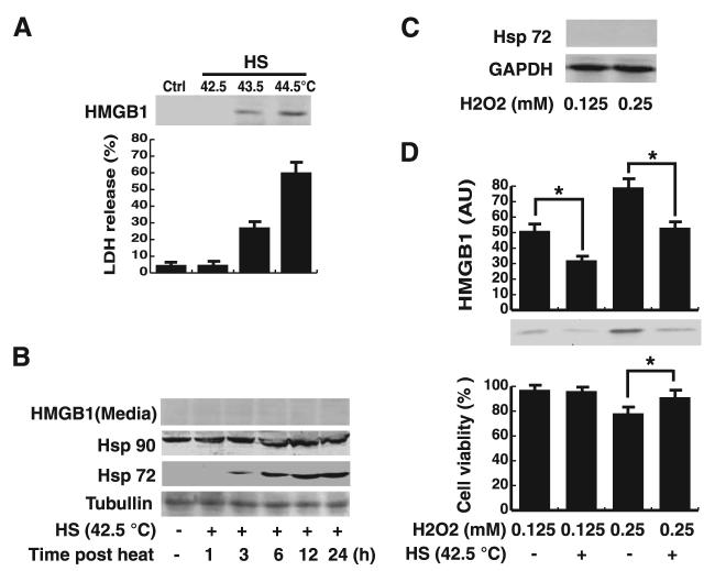
FIGURE 1.
A mild heat shock (HS) pretreatment attenuates oxidative stress (H2O2)-induced HMGB1 release in macrophages cultures. A, Effects of mild or severe heat shock on HMGB1 release. RAW 264.7 cells were subjected to a brief heat shock (1 h) at 42.5°C (mild heat shock), 43.5°C or higher (severe heat shock), after which was returned to 37°C and incubated for additional 12 h. HMGB1 in the culture medium were detected by Western blotting analysis (top). In parallel, the cell viability was determined by LDH release assay (bottom). Ctrl, control cells. Blot is representative of three experiments with similar results. Values are mean ± SEM (n = 3) of three experiments in duplicate. B, Mild HS pretreatment induced Hsp expression. RAW 264.7 cells were subjected to heat shock (42.5°C, 1 h), and cellular levels of Hsp90 and Hsp72 were detected by Western blotting analysis at the indicated time points after heat shock. In parallel, levels of HMGB1 in the culture medium were determined by Western blotting analysis. Tubulin was used as a loading control. Values are representative of three independent experiments with similar results. C, Effects of oxidative stress on Hsp72 expression. RAW 264.7 cells were stimulated with H2O2 at nontoxic (0.125 mM), or low-toxic (0.25 mM) concentrations, and cellular Hsp72 were detected at 12 h poststimulation by Western blotting analysis. GAPDH was used as a loading control. Values are representative of three independent experiments with similar results. D, Mild heat shock pretreatment inhibited H2O2-induced HMGB1 release. After mild heat shock (42.5°C, 1 h), cells were allowed to recover for 12 h at 37°C and then stimulated for 12 h with H2O2 at nontoxic (0.125 mM) or low-toxic (0.25 mM) concentrations. Levels of HMGB1 in the culture medium were determined by Western blotting and expressed (in arbitrary units; AU) as mean ± SEM of three experiments in duplicate. In parallel, the cell viability was determined by MTT assay, and expressed as mean ± SEM (n = 4) of three experiments in duplicate. *, p < 0.05.

