
FIGURE 3.
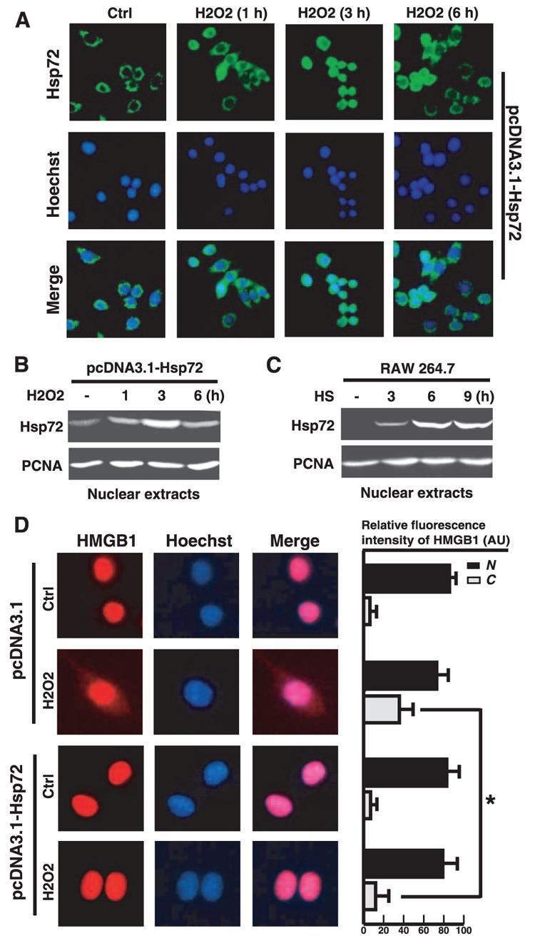
Overexpression of Hsp72 attenuates H2O2-induced HMGB1 cytoplasmic translocation in macrophage cultures. A and B, H2O2 induces transient nuclear translocation of Hsp72 in macrophages cultures. pcDNA3.1-Hsp72-transfected RAW 264.7 cells were stimulated with H2O2 (0.125 mM) for various period of time and examined for Hsp72 subcellular localization by immunocytochemistry (A) or cell fractionation/Western blot (B). Green, Hsp72; blue, nuclei. Original magnification, ×400. A nuclear protein, proliferating cell nuclear Ag, was used as a loading control. C, Heat shock induces Hsp72 expression and nuclear translocation in macrophages cultures. RAW 264.7 cells subject to mild heat shock (42.5°C, 1 h), and nuclear Hsp72 content was determined by Western blotting. D, Overexpression of Hsp72 attenuates H2O2-induced HMGB1 cytoplasmic translocation. RAW 264.7 cells transfected with empty pcDNA3.1 plasmid, or pcDNA3.1-Hsp72 construct were stimulated with H2O2 (0.125 mM) for 12 h, and the subcellular localization of HMGB1 was determined by immunocytochemical analysis (D, left). Relative fluorescence intensity of HMGB1 in the nuclear (N) and cytoplasmic (C) regions of multiple representative cells was assayed using the ImageProPlus software (D, right). Image is representative of three experiments with similar results. Red, HMGB1; blue, nuclei. Original magnification, ×1000. Values are means ± SEM (n = 50) of three experiments in duplicate. *, p < 0.05.