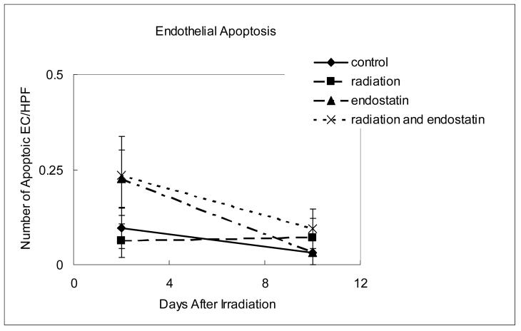
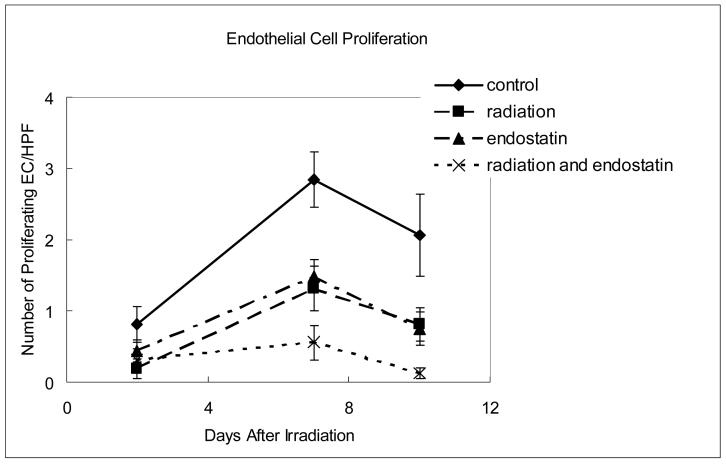
Figure 4.
Time course analyses of endothelial cell apoptosis and proliferation. (a) Endothelial cell apoptosis as determined by CD31-TUNEL double-staining. The number of apoptotic endothelial cells was counted in 7-9 random fields at ×400 magnification. (b) Endothelial cell proliferation as determined by CD31-BrdU double-staining. The number of proliferating endothelial cells was determined in 18-25 random fields at ×200 magnification. (c) Representative staining for CD31 (red fluorescence), BrdU (green fluorescence), or both CD31 and BrdU (yellow fluorescence) in tumor tissue harvested from mice killed 7 days after irradiation.


