
Figure 5. UDPGA binding site is highly conserved in humans.
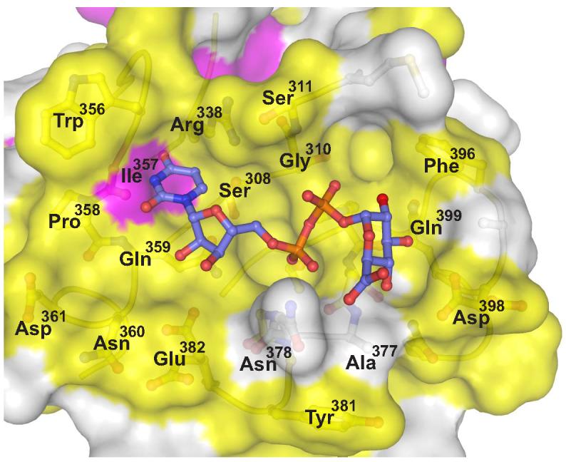
Human UGT sequence conservation (described in Figure 3) is mapped to the molecular surface of 2B7CT. Yellow is invariant, magenta is conserved, and white signifies low conservation. Using the VvGT1 structure as a guide UDPGA was modeled into the 2B7CT structure and is shown in a ball and stick representation.