
FIG. 2.

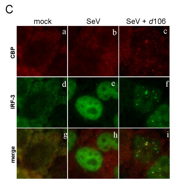
Change of IRF-3 localization in the presence of ICP0. HEC-1-B cells were infected with SeV in the presence or absence of d106. (A) The cells were fixed at 2.5 h post infection and stained with mouse anti-ICP0 and rabbit anti-IRF-3 antibodies with appropriate secondary antibodies. (B) The cells were stained with mouse anti-p300 and rabbit anti-IRF-3 antibodies with appropriate secondary antibodies. (C) The cells were stained with mouse anti-CBP and rabbit anti-IRF-3 antibodies with appropriate secondary antibodies.