
Figure 1.
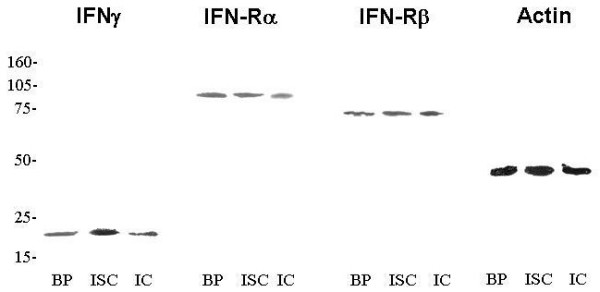
Western blot analysis of IFNγ, IFNγ-Rα, IFNγ-Rβ and α-actin after 15% polyacrilamide gel electrophoresis. BP, benign pathologies. ISC, in situ carcinoma. IC, infiltandrating carcinoma. For each antibody used, a single band – at their corresponding molecular weight – was found: IFNγ (17 kDa), IFNγ-Rα (80 kDa), IFNγ-Rβ (70 kDa) and α-actin (40 kDa).