Figure 5.
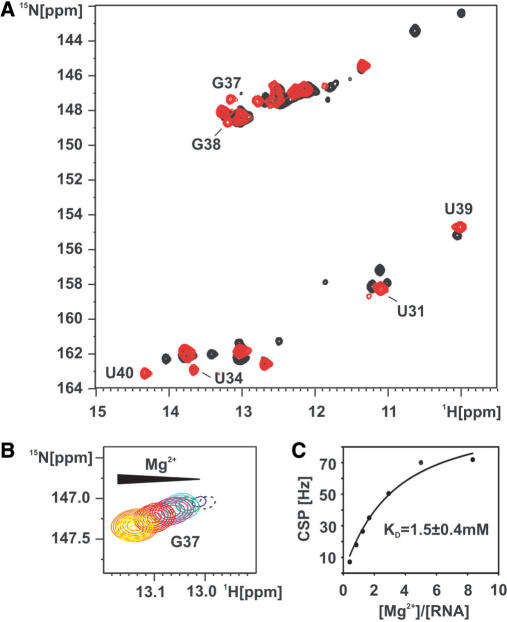
Mg2+ induces folding of the loop–loop interaction in the free form of the adenine-sensing riboswitch RNA. (A) Overlay of the imino region of a 1H,15N-HSQC spectrum of the free (black) and the Mg2+ bound form (red) of the aptamer domain of the adenine-sensing riboswitch RNA. G37 is barely detectable in the free form of the RNA but strongly increases in intensity upon addition of 5 mM Mg2+. (B) 1H,15N-HSQC spectrum of the imino group of G37 upon titration with Mg2+. Mg2+ titration steps are 0 mM (black), 0.25 mM (blue), 0.5 mM (green), 0.75 mM (cyan), 1 mM (purple) 1.75 mM (red), 3 mM (orange) and 5 mM (yellow). The resonance of the G37 imino group at 0mM Mg2+ is shown in dashed contours since it is plotted on a lower level compared to all other titration steps. (C) The chemical shift perturbation (CSP) of the G37 imino resonance is plotted against the ratio [Mg2+]/[RNA] and fitted by non-linear regression (black line). The derived binding constant (KD) of Mg2+ to G37 is 1.5 ± 0.4 mM.

