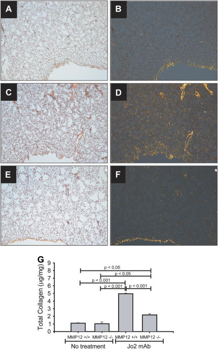
Figure 4.
Collagen response in mmp12−/− mice. Lung sections from mice treated with intratracheal Jo2 mAb for three consecutive days, then killed 21 d after the first instillation (A–F). The tissues were stained with Sirius Red, which stains collagen bright orange under polarized light (right panels). Untreated WT mice have a signal mostly at the pleural surface (A, B). WT mice treated with Jo2 mAb show a marked increase in the orange signal (C, D). In contrast, mmp12−/− mice show markedly less orange signal for collagen (E, F) (×40). Whole collagen content in whole lungs was confirmed using Sircoll collagen assay (G).

