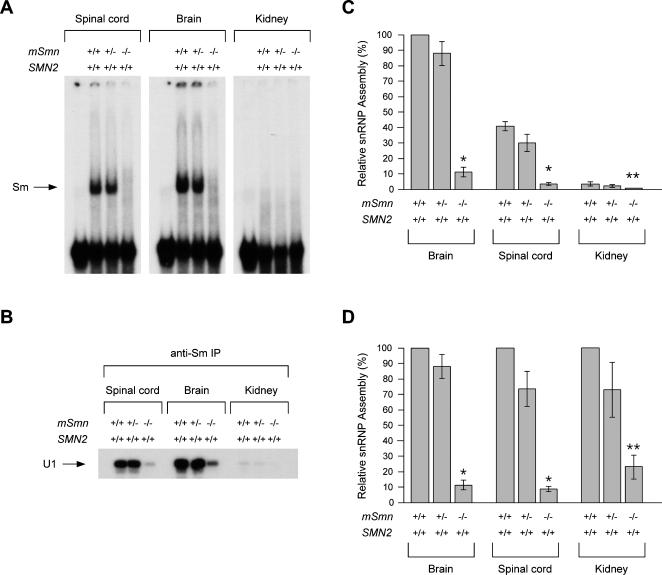
Figure 3. In vitro snRNP assembly activity in tissues of severe SMA mice.
(A) Equal amounts of whole tissue extracts (25 µg) from spinal cord, brain or kidney of normal (SMN2 +/+;mSmn +/+), carrier (SMN2 +/+;mSmn +/−) and severe SMA (SMN2 +/+;mSmn −/−) mice at postnatal day 3 were analyzed in snRNP assembly reactions with radioactive U1 snRNA followed by electrophoresis on native polyacrylamide gels. The position of U1 RNP complexes containing the Sm core is indicated on the left. (B) In vitro snRNP assembly reactions as in (A) were analyzed by immunoprecipitation with anti-Sm (Y12) antibodies followed by electrophoresis on denaturing polyacrylamide gels and autoradiography. (C) Quantification of snRNP assembly activity in whole tissue extracts from normal, carrier and SMA mice. The amount of U1 snRNA immunoprecipitated in snRNP assembly experiments as in (B) was quantified using a STORM 860 Phosphorimager (Molecular Dynamics) and expressed as a percentage of that in brain extracts of normal mice, which is set arbitrarily as 100%. The values from four independent experiments are presented as mean±SEM (*p<0.0001, **p<0.05). (D) For each tissue, the amount of U1 snRNA immunoprecipitated in snRNP assembly experiments with extracts from carrier (SMN2+/+;mSmn+/−) and SMA (SMN2+/+;mSmn−/−) mice is expressed as a percentage of that in the corresponding extract from normal (SMN2+/+;mSmn+/+) mice, which is set arbitrarily as 100%.

