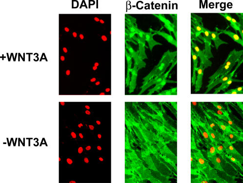
Figure 1. Immunofluorescence analysis comparing ß-Catenin localization in Wnt-treated and mock-treated human dermal fibroblasts.
Immunofluorescence staining shows accumulation of nuclear ß-Catenin in Wnt-treated cells, but no expression in “mock” treated cells (after 4 hrs of Wnt treatment). DAPI was used to stain cell nuclei (red).

