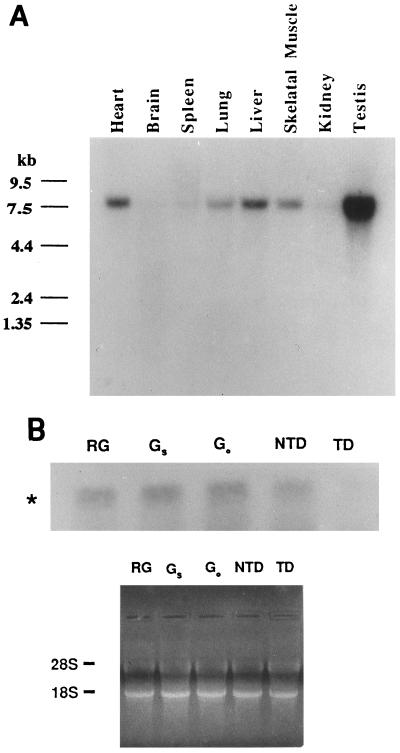
Figure 3.
Tissue distribution of the P2P-R mRNA and its specific repression by terminal adipocyte differentiation. (A) A murine multiple tissue Northern blot (CLONTECH) was analyzed using 32P-labeled random-primed P2P-R cDNA probes under high-stringency conditions. Size markers in kilobases (kb) are shown on the left. (B) Total cellular RNA (20 μg) isolated from growing undifferentiated 3T3T cells (RG), quiescent serum-starved undifferentiated 3T3T cells (Gs), quiescent predifferentiation arrested 3T3T cells (GO/GD), nonterminally differentiated 3T3T adipocytes (NTD), and terminally differentiated 3T3T adipocytes (TD) were hybridized with 32P-labeled random-primed P2P-R cDNA probes under high-stringency conditions. The 8-kb P2P-R mRNA (∗) is shown. A photograph of the ethidium bromide-stained gel before nucleic acid transfer to the nitrocellulose membrane is shown to indicate equivalent amounts of RNA in each lane.

