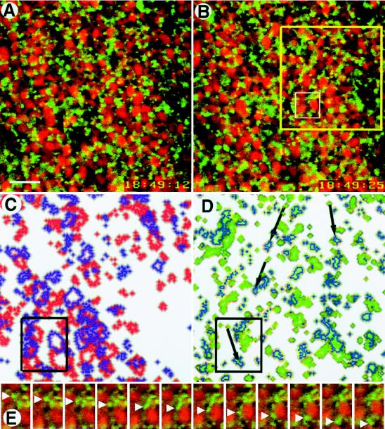
Figure 1.
Rapid movement of DiOC6(3)-labeled organelles in the vegetal pole region of an egg during cortical rotation. Optical sections were collected at a frequency of one per second 4–8 μm inside the vegetal surface and then compiled into a time-lapse video. (A and B) The two images shown here were collected 13 sec apart. The red ovals are nile red-stained yolk platelets; between the time of the first image and the second, they have moved toward the upper left at a velocity of ≈10 μm/min. The smaller green circles are DiOC6(3)-labeled organelles; between the first and second images, most of them have moved toward the upper left (with the yolk platelets), but ≈10% have moved in the opposite direction at ≈30 μm/min. Organelles were continuously tracked through the intervening images. The yellow box (B) outlines the region shown in C and D; the white box in B outlines the region shown in E. (C) Compiled image of a portion of A and B (yellow box, B), at slightly higher magnification, showing change in position of yolk platelets from A (red outlines) to B (purple outlines). (D) Compiled image of a comparable portion of A and B (yellow box, B) at slightly higher magnification showing change in position of DiOC6(3)-labeled organelles from A (green outlines) to B (blue outlines). The black arrows indicate the routes taken by some of the 10% of small organelles that moved rapidly in the opposite direction of yolk platelet movement. The black boxes in C and D correlate with the white box shown in B. (E) Close-ups taken 1 sec apart in the interval between A and B showing a DiOC6(3)-labeled organelle (green, white arrowheads) moving from top to bottom and a nile red-labeled yolk platelet (red) moving from bottom to top. By the 12th image, the small organelle has moved more than twice as far as the yolk platelet (Bar = 10 μm.)

