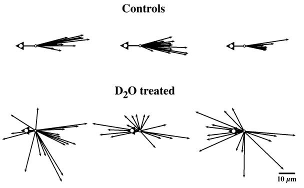
Figure 2.
Direction of organelle displacements 4–8 μm inside the vegetal surface of immobilized eggs undergoing cortical rotation. Arrowheads indicate the direction of yolk platelet movements that, in control eggs, averaged 11.3 μm. In 2H2O-treated eggs, yolk platelet displacement ranged between 0.3 and 12.2 μm, with an average displacement of 5.4 μm (n = 12). Arrows show the directions of rapidly moving DiOC6(3)-labeled organelles. All vectors are brought to a common origin, and the length of the arrows is proportional to the distance traveled by the organelle before it stopped or left the field of view. Only displacements ≥ 3 μm were scored. In control eggs, the rapidly moving organelles are uniformly transported in the opposite direction from the yolk platelets; in 2H2O-treated eggs, their direction of movement is randomized away from the vegetal pole. Randomized movement also was seen in an additional six 2H2O-treated eggs (not shown here). (D2O = 2H2O.)

