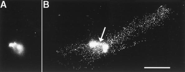
Figure 3.
Movement of fluorescent beads from the vegetal pole to the equatorial region during cortical rotation. (A) A bolus of beads at the vegetal pole before the onset of cortical rotation. (B) A streak of beads in an egg fixed at the end of cortical rotation, when the first cleavage furrow has begun to form. A linear array of beads is seen extending ≈600 μm from the center of the bolus of beads at the vegetal pole (arrow) toward the equator on the side of the egg opposite the sperm entry point, where the future dorsal side usually forms. Fixed eggs were cut in half along the animal–vegetal axis perpendicular to the cleavage furrow, and each half was compressed between two coverslips (to a final thickness of ≈100 μm) to facilitate visualization of the future dorsal and ventral halves. Optical sections were collected at 0.2-μm intervals, starting at the cell surface and moving into the cytoplasm, to visualize the entire transport zone located 4–8 μm from the cell surface. The final image represents a projection of these sections and shows the entire streak of beads. In some eggs (not shown), beads leaked from the injection needle into the deep cytoplasm between the injection site and the vegetal pole. These streaks were easily distinguished from streaks caused by cortical rotation because they were in the half of the egg containing the sperm entry site, were several hundred microns deeper in the cytoplasm, and were more visible from the bisected cytoplasmic side of the egg than from the plasma membrane. (Bar = 200 μm.)

