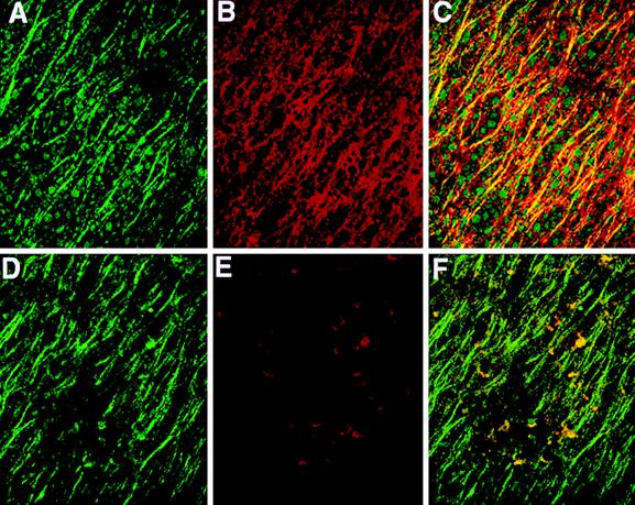
Figure 4.
Colocalization of β-catenin with subcortical microtubules on the future dorsal side of Xenopus eggs at the end of cortical rotation (0.8 of the first cell cycle). Images show labeling with antibodies to β-tubulin and β-catenin (n = 9). (A) Microtubules (green) of the parallel array on the future dorsal side 60–90° from the vegetal pole; (B) linear array of β-catenin (red) in the same region 4–8 μm from the cell surface; (C) colocalization of microtubules and β-catenin (yellow) in this region; (D) microtubules (green) on the future ventral side 60–90° from the vegetal pole; (E) this same (ventral) region shows only small isolated patches of β-catenin; and (F) linear pattern is green rather than yellow (in contrast to C), indicating no colocalization of β-catenin with microtubules on the ventral side. (Bar = 50 μm).

