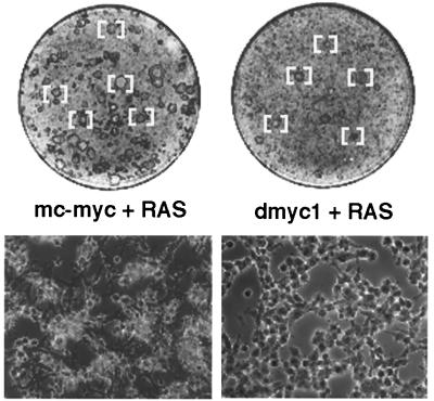
Figure 2.
Gross and microscopic appearance of transformed foci generated in REF experiments presented in Table 1. (Upper) A subset of mc-myc/RAS- or dmyc1/RAS-transformed foci [bracketed] as seen in a Wright–Giemsa-stained monolayer on day 11 post-transfection (experiment 2 from Table 1). All foci were confirmed by microscopic inspection. (Lower) Microscopic appearance of a representative transformed focus under phase contrast photographed on a Zeiss axioscope at 130× magnification.

