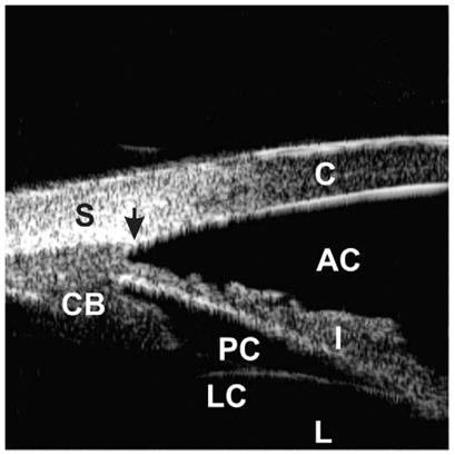
Fig. 1.

Ultrasound biomicroscopic appearance of a normal eye. The cornea (C), sclera (S), anterior chamber (AC), posterior chamber (PC), iris (I), ciliary body (CB), lens capsule (LC), and lens (L) can be identified. The scleral spur (black arrow) is an important landmark to assess the morphologic relationships among the anterior segment structures.
