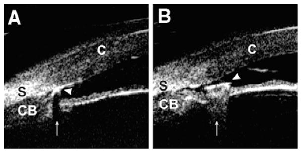
Fig. 12.

Intraocular foreign body. (A) Foreign body (arrow head) with a material that consists of multiple cavities inside (ie, wood and concrete) generates shadowing artifact (arrow) by absorbing ultrasound power. The iris image is masked by shadowing. (B) Hard and dense foreign body (arrow head) (ie, glass and metal) creates comet tail artifact (arrow) owing to multiple internal reflections. The iris image is disrupted by the comet tail artifact. (Adapted from Laroche D, Ishikawa H, Greenfield D, et al. Ultrasound biomicroscopic localization and evaluation of intraocular foreign bodies. Acta Ophthalmol Scand 1998;76(4):491–5; with permission.)
