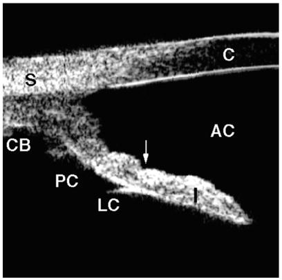
Fig. 8.

Pigment dispersion syndrome. The angle is wide with a concave iris (arrow). Note the extremely wide iridolenticular contact. (Adapted from Breingan PJ, Esaki K, Ishikawa H, et al. Iridolenticular contact decreases following laser iridotomy for pigment dispersion syndrome. Arch Ophthalmol 1999;117(3):325–8; with permission.)
