
Figure 2.
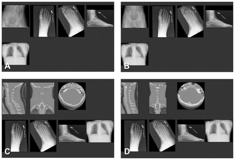
Case 28 as it appeared in thumbnails in: (A) the SOS condition of experiment 1; (B) the non-SOS condition of experiment 1; (C) the SOS condition of experiment 2; and (D) the non-SOS condition of experiment 1. The test fracture was a right navicular fracture visible on the foot (when shown on a 3 megapixel monitor, A–D). The chest examination was normal (A–D). The minor added fracture in experiment 1 was a left acetabular fracture (A). The corresponding pelvic study for the non-SOS condition was a normal pelvis (B). The major fracture in experiment 2 was a C6 anterior subluxation with bilateral facet fracture (C). The corresponding cervical spine CT study for the non-SOS condition was normal (D).