
Figure 3.
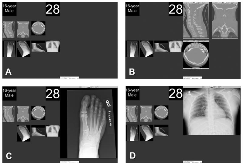
A dual-monitor display of case 28 as it appeared in experiments 2 and 3. Each case was initially presented with patient information, case number, and thumbnail images comprising the complete study on the left monitor (A). Images were displayed, one examination at a time, on the right monitor for diagnostic interpretation (B–D). In the first examination display (B), the three images are sagittal, coronal, and axial image stacks of a cervical spine CT demonstrating a C6 anterior subluxation with bilateral facet fracture. This fracture/subluxation is associated with major morbidity and served as a major added fracture. The next three images are digital radiographs of the foot demonstrating a subtle navicular fracture that served as a test fracture (C). The chest radiograph did not demonstrate a fracture (D).