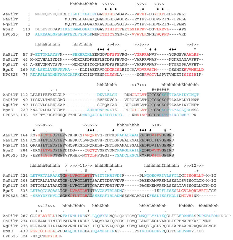
Figure 1.

The sequence and secondary structure of A. aeolicus PilT (AaPilT) aligned with P. aeruginosa PilT (PaPilT), N. gonorrhoeae PilT (NgPilT), V. cholerae EpsE (EpsE) and H. pylori HP0525 (HP0525). Alignments are based on 3-D superpositions, sequence homology, and manual optimization. Observed A. aeolicus PilT α-helices (h) and β-strands (>) are denoted above the sequence, and indicated by blue and red letters, respectively, for the three known structures. Type II/IV secretion ATPase motifs are indicated by shaded boxes: Walker A, Asp Box, Walker B and His Box, in that order. Other symbols mark the CTDn:NTDn+1 interface (diamond), recognition of adenine (plus sign), arginine wire (quotation), other residues presumed to participate in ATP recognition and catalysis (pound sign), and positions of changes in other non-functional PilT variants from this study (dagger). Dots indicate every 10th amino acid in the A. aeolicus PilT sequence. The EpsE zinc-binding tetracysteine motif (CM subdomain) is marked by a carat; the alignment continues after this 40 amino acid insertion. Residues not resolved in the refined structures are typed in grey. EpsE and HP0525 sequences are not shown upstream of the region of structural homology.
