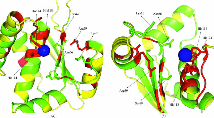
Figure 4.
Cleft structure prepared by PyMol (DeLano, 2002 ▶). The cleft structure is shown in secondary structure colored by conservation score as in Fig. 3 ▶. Conserved residues in the cleft that are possibly involved in function are represented by sticks. Blue sphere is the Ni2+ ion. A side view and a top view are shown in (a) and (b), respectively.

