
Figure 2. Pam3CSK4 induces IRAK-M nuclear export.
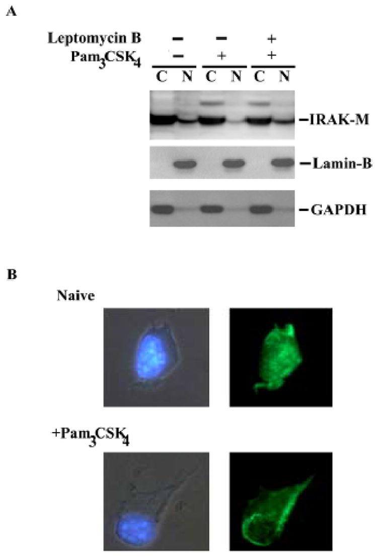
A), Naïve or leptomycin B-pretreated THP-1 cells were subsequently challenged with 100 ng/ml Pam3CSK4 for one hour. Cytoplasmic (C) and nuclear (N) extracts were harvested, separated on SDS-PAGE and blotted with IRAK-M, GAPDH or Lamin-B antibody. GAPDH and Lamin-B are cytoplasmic and nuclear markers, respectively. B), primary murine bone marrow derived macrophages (BMDM) were challenged with 100 ng/ml Pam3CSK4 for one hour. Treated or non-treated cells were fixed, incubated with IRAK-M antibody, and then stained with cy-2 -conjugated anti-Rabbit IgG (right panel). The cells were double stained with DAPI to visualize the nuclei (left panel).