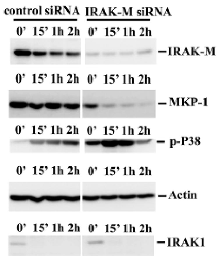
Figure 5. IRAK-M regulates MKP-1 stability in THP-1 cells.

A), THP-1 cells were transfected with control or IRAK-M specific siRNA as described in the materials and methods. Transfected cells were subsequently challenged with 100 ng/ml Pam3CSK4 for various time periods. Equal amounts of total cell lysates were separated on SDS-PAGE, and MKP-1 protein levels were determined through Western blot using anti-MKP-1 antibody. B), Hela-TLR2 (MAT-2) cells were transfected with either empty pcDNA vector or pflag-IRAK-M as described in the Experimental Procedures. Transfected cells were subsequently challenged with 100 ng/ml Pam3CSK4 for various time periods. Equal amounts of cell lysates were separated on SDS-PAGE, and Western blot analyses were performed using anti-MKP-1 antibody. Results were representative of three independent experiments.
