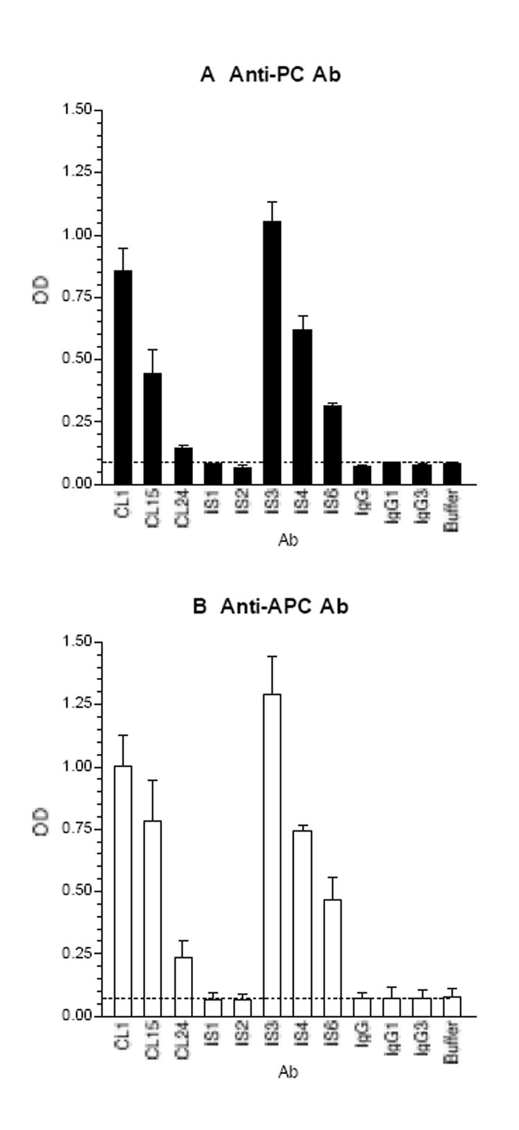
Figure 3.

The thrombin-reactive monoclonal aCL/aPT bind to PC (panel A) and APC (panel B). Microtiter wells were coated with PC or APC, and the test mAb and control IgG were analyzed at 1 μg/ml. IS1 and IS2 are IgG1, and the other mAb are IgG3. Bound IgG was measured and expressed in OD; the means and the ranges are given (n = 2).
