
Fig. 3.
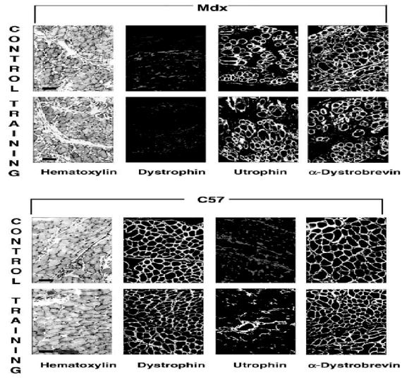
Comparative analysis on cryostat sections from (a) upper panel: trained and control mdx mice, (b) lower panel: trained and control C57BL10 mice. In mdx mice, compared to C57BL10, hematoxylin staining of diaphragm revealed infiltration by interstitial tissue and clear central nuclei distribution in some muscle cells. Immunofluorescence labeling was obtained using specific polyclonal antibodies directed specifically against dystrophin, utrophin and a-dystrobrevin. As expected, utrophin staining was clearly located in the plasma membrane from dystrophin-deficient muscles and dystrophin was absent from mdx muscles. α-Dystrobrevin was present in all muscles. There was no major difference revealed by immunofluorescence between control and training muscles in either mdx or C57BL10 mice.