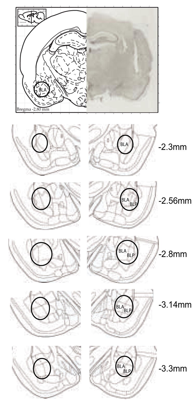
Figure 1. Photomicrograph of a representative cannula placement and schematic representations of injection sites into the BLA at the indicated rostrocaudal planes.

The numbers represent the coordinates from bregma (in millimeters) according to Paxinos and Watson (1998). Injections sites were contained within the black circle. BLA: basolateral amygdaloid nucleus, anterior part; BLP: basolateral amygdaloid nucleus, posterior part.
