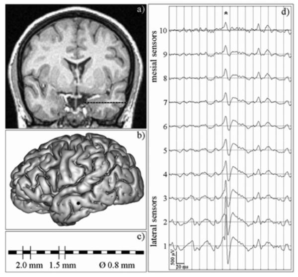
Fig. 3.

An example of SEEG exploration. For simplicity, only one intracerebral electrode is represented. However, during presurgical evaluation, the usual procedure uses 5 to 10 electrodes to explore brain regions that are supposed to be involved in epileptic activity, (a) MRI data of a patient suffering from lateral temporal lobe epilepsy. An intracerebral electrode is implanted in the left middle temporal gyrus, orthogonally to the sagittal plane, (b) Lateral view of the cortex, with surgical entrance point of the electrode in the left middle temporal gyrus. (c) Along each electrode, 10 to 15 cylindrical sensors (length: 2 mm, diameter: 0.8 mm, 1.5 mm apart) record signals from mesial (deep) and from lateral neocortical (middle temporal gyrus, superior temporal gyrus) structures, (d) An example of depth-EEG signals recorded by a 10-sensor intracerebral electrode, from mesial sensors (#7–10) to lateral, i.e. neocortical, sensors (#1–4). Asterisk indicates an interictal epileptic spike with maximal amplitude at lateral sensors and progressive amplitude decrease from lateral to mesial sensors.
