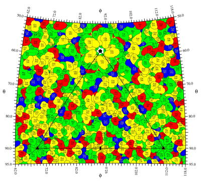
Figure 3.
A spherical roadmap of CPV surface residues. Basic, acid, polar, and hydrophobic residue are colored blue, red, yellow, and green, respectively. A little more than one icosahedral asymmetric unit is shown. The borders of the asymmetric unit are outlined in black, and the icosahedral symmetry axes are labeled with corresponding symbols.

