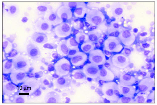
Figure 2.
Photomicrograph of a cytology slide from an aspirate of a cutaneous mass on a 2-year-old dog. The mass was diagnosed as cutaneous histiocytoma. Note the pale cytoplasm and round nuclei typical of this cell type. Image courtesy of Dr. Jeff Sirninger, Louisiana State University. Bar = 10 μm.

