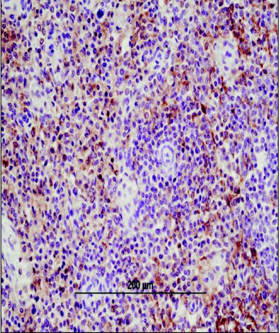
Figure 5.
Photomicrograph of splenic tissue exhibiting diffuse CD18 staining by immunohistochemistry. The CD18 positive cells are identified by their golden-brown staining pattern. CD18 staining is performed on formalin-fixed tissue and although it is a broad leukocyte marker, staining is prominent in histiocytic tumors. Image courtesy of Dr. Tim Morgan, Louisiana State University. Bar = 200 μm.

