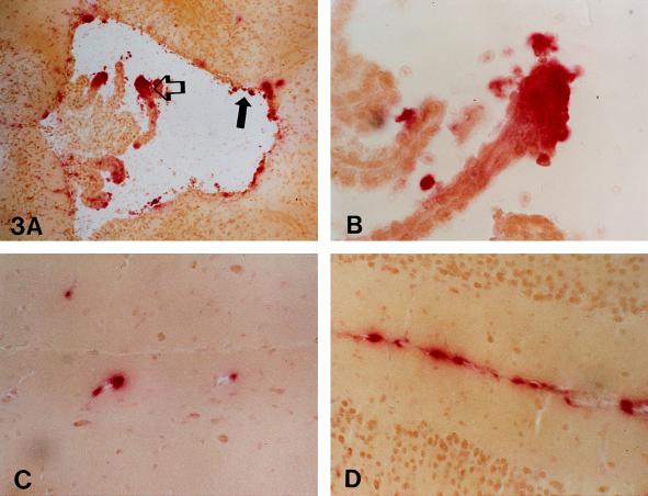
Figure 3.
The ependyma (solid arrow) and choroid lining (open arrow) of the third ventricle (A and B) contained histochemically demonstrable β-glucuronidase activity 15 days after 4.48 × 10⁸ pfu of recombinant adenovirus was injected into the lateral ventricle. In the same mouse, scattered β-glucuronidase-positive cells were also seen in cells associated with vessels (C) in the parenchyma. The meninges (D) contained perivascular and parenchymal cell β-glucuronidase activity. (A, ×50; B, ×210; C and D, ×130.)

